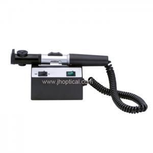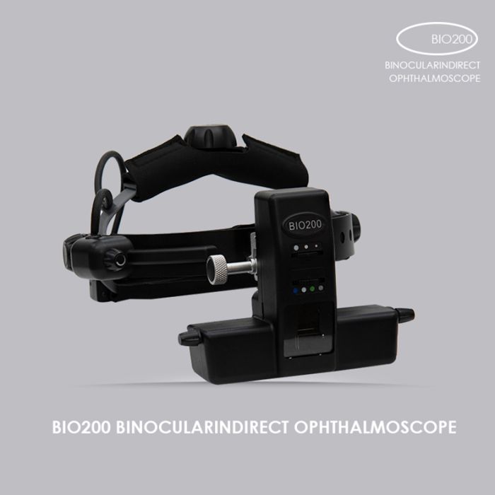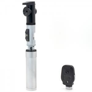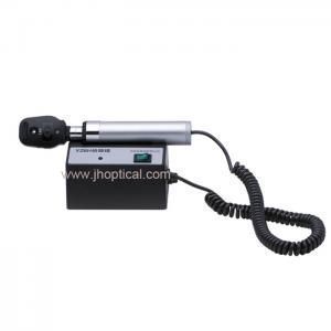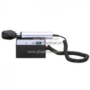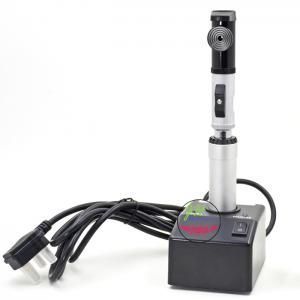
YZ24B Streak retinoscope (halogen lamp)
?
Sign In
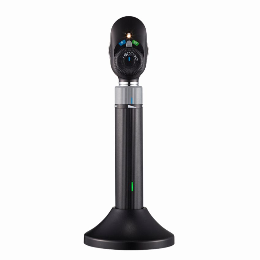
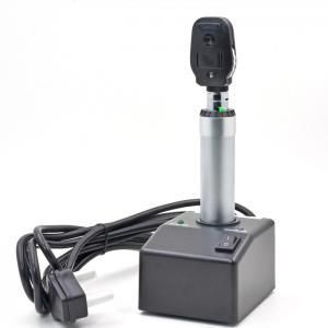
The YZ11E is designed to examine both the external and internal structures of the eye, with a particular focus on the media and the fundus. It is an essential tool for routine eye examinations, diagnostic assessments, and clinical evaluations.
The YZ11E direct ophthalmoscope utilizes a high-quality halogen tungsten light source, delivering reliable and consistent illumination. It features:
The device integrates an illuminating system, an observation system, and viewing lenses that allow the observer to visualize the patient's eye directly—without the formation of an intermediate image. This direct optical path ensures clear, real-time observation for accurate diagnosis.
The YZ11E direct ophthalmoscope is composed of three main parts:
| Parameter | Specification |
|---|---|
| Compensating Lenses (Diopter Compensating Plates, Unit D) | 0, ±1, ±2, ±3, ±4, ±5, ±6, ±8, ±10, +12, ±15, ±20, -25, -35 |
| Diaphragms | Slit, Ophthalmoscope graticules, Wide angle, Intermediate, Macular, Semi-circle |
| Lighting Source | 3.5V 2.8W halogen tungsten bulb |
| Power Adapter Input | 100–240V a.c. 50/60Hz |
| Power Adapter Output | 5V d.c. 1A MAX |
| Internal Battery | 3.7V d.c. 2600mAh Li-ion battery |
| Altitude | ≤3000m |
| Use Environment | Typically used in an indoor darkroom environment |
| Overvoltage Category | II |
| Pollution Degree Classification | 2 |
| Serial Number | Name | Quantity | Remarks |
|---|---|---|---|
| 1 | Optical head | 1 | / |
| 2 | Handle | 1 | / |
| 3 | Power adapter | 1 | / |
| 4 | Spare light bulbs | 1 | / |
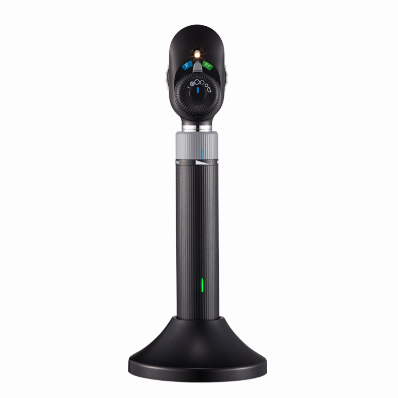
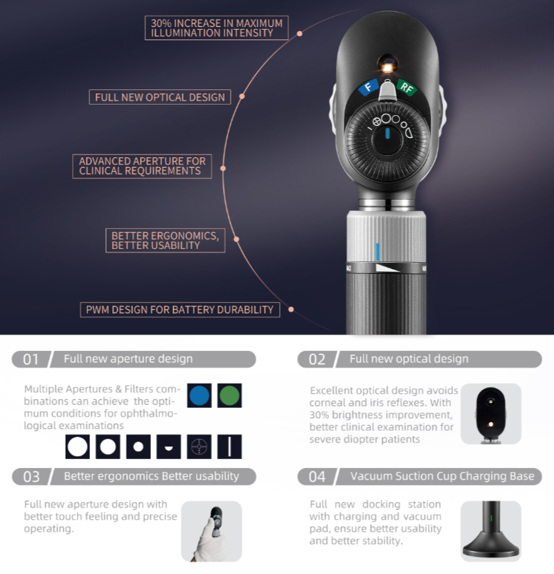
YZ11E New Direct Ophthalmoscope - Key Upgrades
1. Brand-New Optical Structure DesignThe color filter disc and functional diaphragm adopt a layered structure, ensuring versatile observation capabilities for diverse clinical needs.
2. Enhanced Operation ExperienceEvolved precision operation experience for improved handling and control.
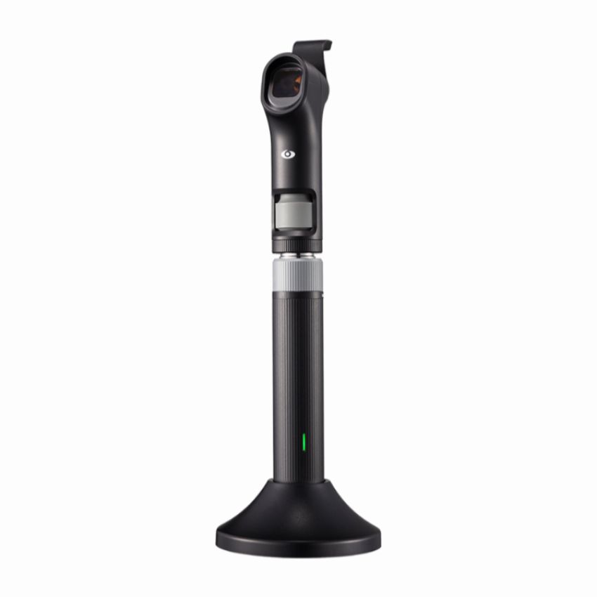
3. New Charging Base DesignDesktop-close center-of-gravity design with negative-pressure suction cup for stable placement; durable charging interface ensures reliable power supply.
4. Upgraded Diaphragm Disc FunctionsLarge, medium, and small spot sizes better match diverse usage habits; precision grid spot and new semi-circular spot are optimized for clinical requirements.
5. Upgraded Optical DesignMaximum illumination intensity increased by 30%, providing clearer fundus observation.
YZ11E - Detailed Feature BreakdownNew Optical Path DesignThe YZ11E features a redesigned layered structure with separate color filter disc and functional diaphragm disc, replacing the older integrated design. This separation ensures more versatile and efficient observation across a wider range of clinical scenarios.Ergonomic Grip Posture AdjustmentThe new dial structure allows the thumb to operate all rotary dials - a significant improvement over the previous model where the thumb operated the power dial and the index finger operated the diaphragm dial. This unified thumb operation significantly improves efficiency and reduces hand fatigue during extended examinations.Significance of Increased Brightness Upper LimitThe 30% boost in maximum brightness is clinically significant in the following scenarios:Opaque Refractive Media - When patients present with corneal edema, cataracts, or vitreous opacities, increased brightness helps penetrate opaque areas to ensure adequate light reaches the retina.Peripheral Retinal Examination - After pupillary dilation (e.g., using tropicamide), higher brightness enables clear observation of the peripheral retina and ora serrata while reducing light scatter interference.Use with Special Filters - When using red-free filters (green filters), which significantly reduce light intensity entering the fundus, the increased brightness compensates to enhance retinal vessel-to-background contrast, aiding detection of hemorrhages and microaneurysms.